
Důkazem vysokého vzdělání je schopnost mluvit o největších věcech nejjednodušším způsobem.
David Hume


Výzkum
Study of fluorescence of doxorubicin in muscle tissue using highly sensitive fluorescence sensing
 Malignant diseases represent 25% of the cause of death and other serious health problems in developed countries. Colorectal cancer, breast cancer, prostate cancer and lung cancer are the most common types of cancer are. On the other hand, mortality caused by this disease gradually decreases and survival time increases due to early diagnosis and effective treatment [1]. Magnetic resonance imaging (MRI), ultrasound (US), positron emission tomography (PET), computed tomography (CT), single-photon emission computed tomography (SPECT) and optical imaging methods belong to the contemporary imaging methods used in diagnostics and monitoring of the therapy [2,3].
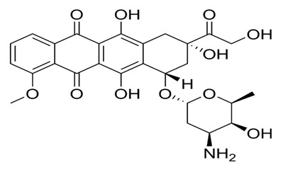
Optical methods are relatively low cost non-ionizing methods based on the specific optical properties. They represent an important tool for non-invasive and objective diagnosis with still better and better resolution [4,5]. It is advantageous to use inherent fluorescent properties of substances and labelled molecules [6]. Fluorescence reflectance imaging (FRI) enables imaging of fluorescent probes in tissues. In this case, the source of radiation and detector are at the same side of the object. Connection of laser and sensitive CCD device together with advanced mathematical models allows sensitive detection and evaluation of fluorescence intensity. Fluorescence-mediated molecular tomography enables reconstruction of three dimensional images of fluorescent probes in the tissue [7-9]. The source of fluorescence for these purposes may be organic fluorophores (fluorescein, rhodamine), biological fluorophores (green fluorescence protein), or quantum dots. On addition, inherent fluorescence of some drugs (doxorubicin, ellipticine) can be used [6,10-12]. In the area of basic research, there is the detection of fluorescence of therapeutics beneficial particularly in the development of targeted therapy and control of drugs targeting into place affected by tumour tissue. Doxorubicin is a highly effective and widely used anthracycline antibiotic, important antineoplastic drug intercalating DNA and causing oxidation stress that is used to treat leukaemia and solid tumours [13-19]. However, its application is limited by high cardiotoxicity, so it is necessary to monitor the applied dose [11,17].
Stationary techniques, such as spectrophotometric methods [20] including fluorimetry [21-23], but also separation methods, such as high performance liquid chromatography [24-28] and capillary electrophoresis [29-32], may be used for in vitro characterization and pharmacological evaluation of doxorubicin. In vivo studying of interactions and distribution of doxorubicin can be performed in a microscale using microscopic techniques in different arrangements as laser scanning microscopy [33-35], fluorescence life time microscopy [14] and scanning electron microscopy [36]. At the macroscale level, high-frequency ultrasound imaging [37], PET imaging [38] and fluorescence imaging using quantum dots [39] have been used for the monitoring doxorubicin or doxorubicin-modified nanoparticles in tissues.
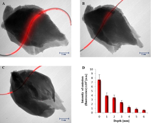
The aim of this work was to study the fluorescent properties of doxorubicin and to study the behaviour of doxorubicin diluted to different concentrations in water or methanol. In addition, doxorubicin was injected into muscle tissue to monitor its behaviour and to detect its fluorescence (emission) at different depths.
Malignant diseases represent 25% of the cause of death and other serious health problems in developed countries. Colorectal cancer, breast cancer, prostate cancer and lung cancer are the most common types of cancer are. On the other hand, mortality caused by this disease gradually decreases and survival time increases due to early diagnosis and effective treatment [1]. Magnetic resonance imaging (MRI), ultrasound (US), positron emission tomography (PET), computed tomography (CT), single-photon emission computed tomography (SPECT) and optical imaging methods belong to the contemporary imaging methods used in diagnostics and monitoring of the therapy [2,3].
Optical methods are relatively low cost non-ionizing methods based on the specific optical properties. They represent an important tool for non-invasive and objective diagnosis with still better and better resolution [4,5]. It is advantageous to use inherent fluorescent properties of substances and labelled molecules [6]. Fluorescence reflectance imaging (FRI) enables imaging of fluorescent probes in tissues. In this case, the source of radiation and detector are at the same side of the object. Connection of laser and sensitive CCD device together with advanced mathematical models allows sensitive detection and evaluation of fluorescence intensity. Fluorescence-mediated molecular tomography enables reconstruction of three dimensional images of fluorescent probes in the tissue [7-9]. The source of fluorescence for these purposes may be organic fluorophores (fluorescein, rhodamine), biological fluorophores (green fluorescence protein), or quantum dots. On addition, inherent fluorescence of some drugs (doxorubicin, ellipticine) can be used [6,10-12]. In the area of basic research, there is the detection of fluorescence of therapeutics beneficial particularly in the development of targeted therapy and control of drugs targeting into place affected by tumour tissue. Doxorubicin is a highly effective and widely used anthracycline antibiotic, important antineoplastic drug intercalating DNA and causing oxidation stress that is used to treat leukaemia and solid tumours [13-19]. However, its application is limited by high cardiotoxicity, so it is necessary to monitor the applied dose [11,17].
Stationary techniques, such as spectrophotometric methods [20] including fluorimetry [21-23], but also separation methods, such as high performance liquid chromatography [24-28] and capillary electrophoresis [29-32], may be used for in vitro characterization and pharmacological evaluation of doxorubicin. In vivo studying of interactions and distribution of doxorubicin can be performed in a microscale using microscopic techniques in different arrangements as laser scanning microscopy [33-35], fluorescence life time microscopy [14] and scanning electron microscopy [36]. At the macroscale level, high-frequency ultrasound imaging [37], PET imaging [38] and fluorescence imaging using quantum dots [39] have been used for the monitoring doxorubicin or doxorubicin-modified nanoparticles in tissues.
The aim of this work was to study the fluorescent properties of doxorubicin and to study the behaviour of doxorubicin diluted to different concentrations in water or methanol. In addition, doxorubicin was injected into muscle tissue to monitor its behaviour and to detect its fluorescence (emission) at different depths.
 Acknowledgements
Financial support from the following projects PGS09_2012 and NANOLABSYS CZ.1.07/2.3.00/20.0148 is highly acknowledged.
Plakáty k výzkumným směrům
Dokumenty pro VaV aktivity
Výzkumný záměr
Hodnocení výzkumných aktivit
Archív
24— 2013
23— 2013
22— 2013
21— 2013
20— 2013
19— 2013
18— 2013
17— 2013
16— 2013
15— 2013
14— 2013
 | Zemědělská 1/1665 613 00 Brno Budova D | Tel.: +420 545 133 350 Fax.: +420 545 212 044 |  |
 |