
Účelem vzdělání není zaplnit mysl, ale otevřít ji. Čím více poznatků si osvojíme, tím víc si uvědomíme, co ještě neznáme.


Výzkum
Microfluidic chip coupled with modified paramagnetic particles for sarcosine isolation in urine
 Genome-wide association studies (GWAS) revealed links between genetic variance and predisposition to disease. With the advent of modern 'omics-technologies', GWAS can now identify the genetic factors that influence intermediate traits on pathways to disease, such as blood concentrations of carbohydrates, lipids, amino acids, and secondary metabolites, hormones and signal molecules [1-4]. Increasing interest about sarcosine as of the secondary metabolites is dated from year 2009, when Sreekumar et al. published their study, which provided new information about possible utilization of sarcosine as a potential prostate carcinoma marker [5]. Sarcosine is CH3NHCH2COOH formulated as a natural, colourless, non-toxic and solid substance well soluble in water. Sarcosine, also N-methylglycine is methyl derivate of glycine, created in organism as an intermediate metabolic products generated during conversion of choline to glycine [6]. Although the connection of sarcosine with amino acids metabolism and methylation processes of prostate tumours progression was described [7-9] as well as its potential in a diagnosis of early stages of prostate tumours [10, 11], usage as a marker is still under discussions, because there can be found some publications that disproved link between sarcosine and prostate tumours [12]. On the other hand, sarcosine is not present in urine of health people, therefore, false positivity is minimized [13, 14]. Recently, numerous methods are developed for sarcosine detection. Most of them are based on different types of chromatography or capillary electrophoresis (CE) or microfluidic chips [15-19] coupled mainly with mass spectrometry [7, 20-28]. There is also possibility to couple modern separation methods with electrochemical detectors, which have low operating costs, the possibility of miniaturization and high sensitivity [29, 30]. Direct electrochemistry using modified glassy carbon electrode was applied by Zhou et al. for sarcosine oxidase detection and detection limits were estimated as 1 µM [31].
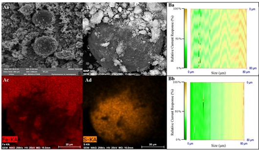
In our study we decided to use ion exchange chromatography with post-column ninhydrin derivatization and VIS detector for sarcosine separation and detection, which minimizes the sample pre-treatment requirements [13]. Major part of the study consisted of sarcosine isolation by Dowex 50WX4-400 microbeads with SO3- functional groups. Surface was modified by paramagnetic particles (PMPs), which are formed by Fe2O3 nanoparticles. Magnetic particles can combine two selective processes in bioanalysis: the specific binding of analytes to the particle surface based on molecular recognition and the specific isolation of magnetic objects from complex sample mixtures [32]. Possibility of MPs surface modification and thereby an elimination of undesirable biomolecules adsorption is the biggest advantage of their utilization [33, 34]. The obtained partially modified surface of our Dowex magnetic particles was characterized by electron microscopy. Application of nanoparticles for isolation of sarcosine was carried out in reaction cell fabricated using 3D printer.
Fabrication of the reaction cell in microchip
Genome-wide association studies (GWAS) revealed links between genetic variance and predisposition to disease. With the advent of modern 'omics-technologies', GWAS can now identify the genetic factors that influence intermediate traits on pathways to disease, such as blood concentrations of carbohydrates, lipids, amino acids, and secondary metabolites, hormones and signal molecules [1-4]. Increasing interest about sarcosine as of the secondary metabolites is dated from year 2009, when Sreekumar et al. published their study, which provided new information about possible utilization of sarcosine as a potential prostate carcinoma marker [5]. Sarcosine is CH3NHCH2COOH formulated as a natural, colourless, non-toxic and solid substance well soluble in water. Sarcosine, also N-methylglycine is methyl derivate of glycine, created in organism as an intermediate metabolic products generated during conversion of choline to glycine [6]. Although the connection of sarcosine with amino acids metabolism and methylation processes of prostate tumours progression was described [7-9] as well as its potential in a diagnosis of early stages of prostate tumours [10, 11], usage as a marker is still under discussions, because there can be found some publications that disproved link between sarcosine and prostate tumours [12]. On the other hand, sarcosine is not present in urine of health people, therefore, false positivity is minimized [13, 14]. Recently, numerous methods are developed for sarcosine detection. Most of them are based on different types of chromatography or capillary electrophoresis (CE) or microfluidic chips [15-19] coupled mainly with mass spectrometry [7, 20-28]. There is also possibility to couple modern separation methods with electrochemical detectors, which have low operating costs, the possibility of miniaturization and high sensitivity [29, 30]. Direct electrochemistry using modified glassy carbon electrode was applied by Zhou et al. for sarcosine oxidase detection and detection limits were estimated as 1 µM [31].
In our study we decided to use ion exchange chromatography with post-column ninhydrin derivatization and VIS detector for sarcosine separation and detection, which minimizes the sample pre-treatment requirements [13]. Major part of the study consisted of sarcosine isolation by Dowex 50WX4-400 microbeads with SO3- functional groups. Surface was modified by paramagnetic particles (PMPs), which are formed by Fe2O3 nanoparticles. Magnetic particles can combine two selective processes in bioanalysis: the specific binding of analytes to the particle surface based on molecular recognition and the specific isolation of magnetic objects from complex sample mixtures [32]. Possibility of MPs surface modification and thereby an elimination of undesirable biomolecules adsorption is the biggest advantage of their utilization [33, 34]. The obtained partially modified surface of our Dowex magnetic particles was characterized by electron microscopy. Application of nanoparticles for isolation of sarcosine was carried out in reaction cell fabricated using 3D printer.
Fabrication of the reaction cell in microchip
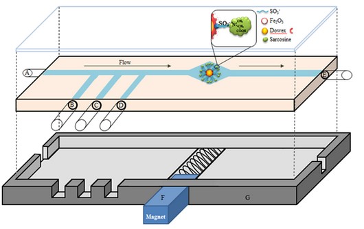 Reaction cell was designed in Blender software (NetFabb Studio, USA) and manufactured under laboratory conditions using 3D printer Easy 3D Maker (3DFactories, Straznice, Czech Republic). The printing material was biodegradable polymer poly-lactic acid (PLA). A distinct advantage of using polymers is the fact that materials with specific chemical and physical properties (such as optical transparency, chemical resistance, stiffness, critical surface tension, etc.) can be selected specifically for a target application [39]. The time of printing was 30 minutes while the density of the matrix was set on 100 %. The pad for the target was tempered on 35 °C.
Nanoparticles – antimicrobial agents
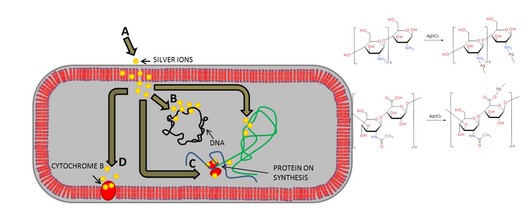 The complexes of polymeric substances (hyaluronic acid and chitosan) with silver nitrate or silver phosphate nanoparticles were created, and their effects on gram-positive bacterial culture of Staphylococcus aureus were monitored. Stages of formation of complexes of silver nitrate and silver phosphate nanoparticles with polymeric compounds were characterized using electrochemical and spectrophotometric methods. Furthermore, the antimicrobial activity of complexes was determined using the methods of determination of growth curves and zones of inhibition.
Plakáty k výzkumným směrům
Dokumenty pro VaV aktivity
Výzkumný záměr
Hodnocení výzkumných aktivit
Archív
24— 2013
23— 2013
22— 2013
21— 2013
20— 2013
19— 2013
18— 2013
17— 2013
16— 2013
15— 2013
14— 2013
 | Zemědělská 1/1665 613 00 Brno Budova D | Tel.: +420 545 133 350 Fax.: +420 545 212 044 |  |
 |