Application of quantum dots into chicken embryos
Iva Blazkova, Marie Konecna, Renata Kensova, Vojtech Adam, Rene Kizek,
Quantum dots (QDs) are small semiconductor nanoparticles with great optical properties. Their behaviour enables the usage of QDs in in vitro and in vivo experiments and they are promising tools in disease treatment and targeted therapy. The limitation of their usage is the toxicity. Quantum dots consist of different metals, which have various effects on the health. To decrease their toxicity, different surface coatings are used. The effect of QDs on the organism can be tested on chicken embryos. Chicken embryos represent great model for QDs toxicity studies, because there is no need of any permission for the work with embryos and the experiments are low cost and fast.

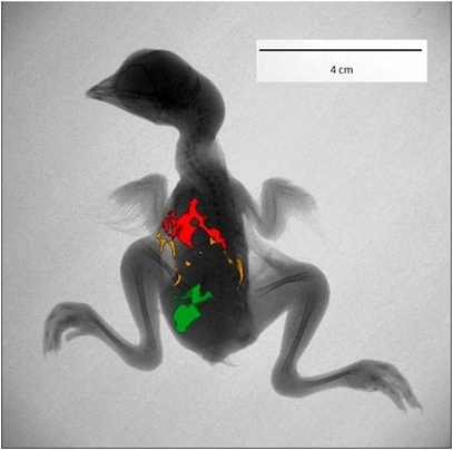
Fig.1: Different CdTe QDs applied into the chicken embryo (18th developmental day) detected by In vivo Xtreme by Carestream (Rochester, NY, USA). The fluorescence of three different QDs detected with excitation filter: 480 nm and emission filter: 535 nm (CdTe green), 600 nm (CdTe yellow) and 700 nm (CdTe red). The fluorescence images were overlayed with x-ray image.
1. Gormus, U. THE RELATIONSHIP OF EMBRYOGENESIS AND CARCINOGENESIS. Nobel Medicus. 2011, 7, 5-9.
2. Giannaccini, M.; Cuschieri, A.; Dente, L.; Raffa, V. Non-mammalian vertebrate embryos as models in nanomedicine. Nanomedicine-Nanotechnology Biology and Medicine. 2014, 10, 703-719.
3. Ivarie, R. Avian transgenesis: progress towards the promise. Trends in Biotechnology. 2003, 21, 14-19.
4. Blazkova, I.; Nguyen, V.H.; Kominkova, M.; Konecna, R.; Chudobova, D.; Krejcova, L.; Kopel, P.; Hynek, D.; Zitka, O.; Beklova, M.; Adam, V.; Kizek, R. Fullerene as a transporter for doxorubicin investigated by analytical methods and in vivo imaging. Electrophoresis. 2014, 35, 1040-1049.
5. Wang, Y.C.; Hu, R.; Lin, G.M.; Roy, I.; Yong, K.T. Functionalized Quantum Dots for Biosensing and Bioimaging and Concerns on Toxicity. Acs Applied Materials & Interfaces. 2013, 5, 2786-2799.
6. Cooper, J.K.; Franco, A.M.; Gul, S.; Corrado, C.; Zhang, J.Z. Characterization of Primary Amine Capped CdSe, ZnSe, and ZnS Quantum Dots by FT-IR: Determination of Surface Bonding Interaction and Identification of Selective Desorption. Langmuir. 2011, 27, 8486-8493.
7. Gomes, S.A.O.; Vieira, C.S.; Almeida, D.B.; Santos-Mallet, J.R.; Menna-Barreto, R.F.S.; Cesar, C.L.; Feder, D. CdTe and CdSe Quantum Dots Cytotoxicity: A Comparative Study on Microorganisms. Sensors. 2011, 11, 11664-11678.
8. Sobhana, S.S.L.; Devi, M.V.; Sastry, T.P.; Mandal, A.B. CdS quantum dots for measurement of the size-dependent optical properties of thiol capping. Journal of Nanoparticle Research. 2011, 13, 1747-1757.
9. Geszke-Moritz, M.; Moritz, M. Quantum dots as versatile probes in medical sciences: Synthesis, modification and properties. Materials Science & Engineering C-Materials for Biological Applications. 2013, 33, 1008-1021.
10. Sobrova, P.; Blazkova, I.; Chomoucka, J.; Drbohlavova, J.; Vaculovicova, M.; Kopel, P.; Hubalek, J.; Kizek, R.; Adam, V. Quantum dots and prion proteins: Is this a new challenge for neurodegenerative diseases imaging? Prion. 2013, 7, 349-358.
11. Tmejova, K.; Hynek, D.; Kopel, P.; Krizkova, S.; Blazkova, I.; Trnkova, L.; Adam, V.; Kizek, R. Study of metallothionein-quantum dots interactions. Colloid Surf. B-Biointerfaces 2014, in press.
12. Ryvolova, M.; Chomoucka, J.; Janu, L.; Drbohlavova, J.; Adam, V.; Hubalek, J.; Kizek, R. Biotin-modified glutathione as a functionalized coating for bioconjugation of CdTe based quantum dots. Electrophoresis. 2011, 32, 1619-1622.
13. Krejcova, L.; Hynek, D.; Kopel, P.; Merlos, M.A.R.; Tmejova, K.; Trnkova, L.; Adam, V.; Hubalek, J.; Kizek, R. Quantum dots for electrochemical labelling of neuramidinase genes of H5N1, H1N1 and H3N2 influenza. Int. J. Electrochem. Sci. 2013, 8, 4457-4471.
14. Bertini, I.; Cavallaro, G. Metals in the "omics" world: copper homeostasis and cytochrome c oxidase assembly in a new light. Journal of Biological Inorganic Chemistry. 2008, 13, 3-14.
15. Lin, G.M.; Ding, Z.C.; Hu, R.; Wang, X.M.; Chen, Q.; Zhu, X.M.; Liu, K.; Liang, J.H.; Lu, F.Q.; Lei, D.L.; Xu, G.X.; Yong, K.T. Cytotoxicity and immune response of CdSe/ZnS Quantum dots towards a murine macrophage cell line. Rsc Advances. 2014, 4, 5792-5797.
16. Mas, A.; Arola, L. CADMIUM AND LEAD TOXICITY EFFECTS ON ZINC, COPPER, NICKEL AND IRON DISTRIBUTION IN THE DEVELOPING CHICK-EMBRYO. Comparative Biochemistry and Physiology C-Pharmacology Toxicology & Endocrinology. 1985, 80, 185-188.
17. Yamamoto, F.Y.; Neto, F.F.; Freitas, P.F.; Ribeiro, C.A.O.; Ortolani-Machado, C.F. Cadmium effects on early development of chick embryos. Environmental Toxicology and Pharmacology. 2012, 34, 548-555.
18. Thompson, J.; Wong, L.; Lau, P.S.; Bannigan, J. Adherens junction breakdown in the periderm following cadmium administration in the chick embryo: distribution of cadherins and associated molecules. Reproductive Toxicology. 2008, 25, 39-46.
19. Thompson, J.; Bannigan, J. Cadmium: Toxic effects on the reproductive system and the embryo. Reproductive Toxicology. 2008, 25, 304-315.
20. Pavlak, K.; Dzugan, M.; Wojtysiak, D.; Lis, M.; Niedziolka, J. Effect of in ovo injection of cadmium on chicken embryo heart African Journal of Agricultural Research. 2013, 1534-1539.
21. Thompson, J.; Doi, T.; Power, E.; Balasubramanian, I.; Puri, P.; Bannigan, J. Evidence against a direct role for oxidative stress in cadmium-induced axial malformation in the chick embryo. Toxicology and Applied Pharmacology. 2010, 243, 390-398.
22. Leon-Buitimea, A.; Rodriguez-Fragoso, P.; Reyes-Esparza, J.A.; Rodriguez-Fragoso, L. Synthesis, characterization and toxicological evaluation of maltodextrin capped cadmium sulfide nanoparticles in human cell lines and chicken embryos. Faseb Journal. 2013, 27.
23. Ribatti, D. Chicken Chorioallantoic Membrane Angiogenesis Model, in: X. Peng and M. Antonyak (Eds.), Cardiovascular Development: Methods and Protocols, Humana Press Inc, Totowa, 2012, pp. 47-57.
24. Jedelska, J.; Strehlow, B.; Bakowsky, U.; Aigner, A.; Hobel, S.; Bette, M.; Roessler, M.; Franke, N.; Teymoortash, A.; Werner, J.A.; Eivazi, B.; Mandic, R. The Chorioallantoic Membrane Assay Is a Promising Ex Vivo Model System for the Study of Vascular Anomalies. In Vivo. 2013, 27, 701-705.
25. Smith, J.D.; Fisher, G.W.; Waggoner, A.S.; Campbell, P.G. The use of quantum dots for analysis of chick CAM vasculature. Microvascular Research. 2007, 73, 75-83.
J.Met.Nano:
volume-1, issue-3
- Personal and professional representation of the nanolabsys project
- Administration and information system of the project
- Microwave preparation of carbon quantum dots with different surface modification
- Cell lines as a model system for quantum dots applications
- Application of quantum dots into chicken embryos
- The influence of zinc to living organisms
- The influence of cadmium to living organisms
- The influence of lead to living organisms
- The influence of mercury to living organisms
- Monitoring of metallothionein levels in biological organism exposed to the metal elements and compounds
- The ratio of GSH/GSSG in biological organisms
 PDF
PDF