World Cancer Day in the Laboratory of Metallomics and Nanotechnologies at Mendel University in Brno
Ondřej Zítka, Zbyněk Heger, Sylvie Skaličková and René Kizek
Laboratory of metallomics and nanotechnologies attended the important event of traditional World cancer day (WCD) which was held on 4. February 2015. On this occasion were prepared a series of lectures aimed for introduction of nanotechnology research as an improvement for cancer diagnosis in early stages, treatment and progressive ways of healing the oncologic patients by gene therapy or rapid cancer diagnosis
Program of WCD 2015 was dedicated to ten researchers from the Laboratory of metallomics and nanotechnologies and their lectures (Fig. 1). The whole series of lectures were introduced by information of the League against cancer Prague activities and regular actions also organized under its auspices on the Laboratory of metalomics and nanotechnology ground. For interest was introduced the cooperation of both subjects which has been continued on the project level since 2006. The connection of some viruses and tumor diseases has been not fully understood yet, so the various relations between malignant progression and presence of SV40 (simian vacuolating virus 40) viral DNA, distributed between 1955 and 1963 in poliomyelitis vaccine as a contamination, were discussed. The ability of virus interference with the function of tumor suppressor p53 was described in laboratory animal tests; however the connections in human have not been clearly described yet.
The main part of lectures was given to nanoLaboratory of metallomics and nanotechnologies attended the important event of traditional World cancer day (WCD) which was held on 4. February 2015. On this occasion were prepared a series of lectures aimed for introduction of nanotechnology research as an improvement for cancer diagnosis in early stages, treatment and progressive ways of healing the oncologic patients by gene therapy or rapid cancer diagnosis Laboratory Report 65 eliminate the toxicity and side effects of cytostatics used in the treatment. In the case of gene therapy based on replacing the defective genes or eliminating its ability to encode a target protein, are employed similar nanotechnologies. Although, the idea of gene therapy is due to the ethical issues complicating acceleration to the clinical applications, the Gendicin (gene which encoded wt-p53 in recombinant adenoviral vector) have been already used successfully for the treatment of malignant cancer squamous cell carcinoma of head and neck in China. Another obstacle that complicates practical application of gene therapy is the need to use viral vectors for gene integration into genome. Although, viruses are recombinantly attenuated, the undesirable massive immune response threatens in some cases. Separate category of nanomaterials, the carbon nanotubes and their modification by drugs or targeting ligands, show a huge application potential. In this case, the nanotoxicologic studies confirm the biocompatability and bioavailability of carbon materials. In vivo and in vitro experiments demonstrate the carbon based carriers improve the passive intake of drugs by cancer cells, while the modification by targeting ligand rapidly increase the elimination of development of different tumor types. The unique properties of nanomaterials could be used not only for drug delivery, but for earlier diagnostic by targeted tumor imaging, both of fluorescence measurement or magnetic resonance where the nanomaterials are employed as contrast agents. In the field of fluorescence are highly discussed semiconductor crystals – quantum dots (QDs) due its higher quantum yield and the possibility of their modification. The main disadvantage of QDs is potential toxicity depending on the forming material, however it has been experimentally demonstrated the toxicity of QDs is negligible in comparison with the presence of the free metal ions. Moreover, the carbon quantum dots especially developed with less toxicity due to toxic metal elimination. Further lectures discussed especially early diagnosis of cancer with the use of biomarkers. Although, the body fluids are the complex matrixes and most of known biomarkers are present in concentrations of the order of less than other matrix compounds, it is necessary to perform a separation step before analysis. The modern analytical methods for biomarkers analysis allow huge possibilities of high resolution and sensitive detection. One of the interesting biomarker is metal-binding protein; metallothionein (MT), its alterations have been identified in a wide range of malignancies. Several studies have been proven the connection of MT into a number of physiological processes such as metal homeostasis and protecting the body against oxidative stress. The main role of MT is the storage and zinc transport thereby contributing to the genes involved to apoptosis, cell proliferation and cellular division. It is therefore not surprising; the level of MT could be used as a supporting tool in diagnostics, prognostics of state of health during and after oncologic treatment.
The word cancer day 2015, held on the ground of Laboratory of metallomics and nanotechnologies, provided scientific and educational Figure 1: Series of lectures given by Laboratory of metallomics and nanotechnologies researchers within WCD 2015 Zitka et al. 66 program coupled with fruitful discussions with all interested researchers. Th e conference fulfi lled the mission to share the information about cancer treatment and early diagnostics using modern analytical methods and interesting biomolecules.
Návštěva bruker axs GMBH Karlsruhe
Zbyněk Heger, Jan Zítka, René Kizek
Cílem pracovní cesty bylo zmapování aplikačních možností různých typů mikroskopů atomárních sil (dále AFM - atomic force microscopy), nabízených firmou Bruker Corp. s ohledem na potencionální kupní zájem. Pracovní cesta byla plánována na dva dny cestovní (18. a 21. 8.) a dva dny pracovní (19. a 20. 8.), strávené v showroomech firmy s danými přístroji, a to především s ohledem na velký počet plánovaných vzorků, které byly dovezeny z Laboratoří Metalomiky a Nanotechnologií Mendelovy univerzity. Vzhledem k biomedicínskému zaměření Laboratoře Metalomiky a Nanotechnologií byly veškeré další práce prováděny právě na přístroji FastScan, který oproti ostatním nabízí rapidně sníženou dobu skenování vzorků se zachováním výborné citlivosti. Jeho výhodou je také protektivní obal, fungující jako Faradayova klec s termo- a akustickou izolací, zvyšující kvalitu výsledného skenu.
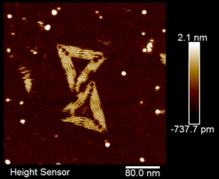 Obr. 1: AFM mikrofotografie DNA origami pořízená na přístroji Dimension Icon (Bruker, Billerica, MA, USA). Autoři děkují Dr. Hartmutu Stadlerovi za perfektní technickou asistenci při získávání tohoto snímku na přístroji firmy Bruker AXS GMBH.
Výsledkem dvou pracovních dnů byla celá řada snímků (přivezené vzorky a DNA origami), získaných pro účely Laboratoře Metalomiky a Nanotechnologií a mini-experiment s pevností a adhezí bakteriofága lambda. Důležitou části setkání bylo také technické představení přístroje, ve kterém byla ukazována běžná údržba, nanášení vzorků na různé typy imobilizačních povrchů (mica, grafit) či výměna kantileveru s mikrojehlou. V rámci softwarové části byla předvedena konfigurace laseru a optické části přístroje a také možnosti různých skenovacích módů a popsán běžný troubleshooting. Během pracovních dnů byly hojně diskutovány výhody a nevýhody jednotlivých přístrojů pro různé biologické, ale i chemické aplikace.
38th International Symposium on Capillary Chromatography (ISCC) and 11th GCxGC Symposium
Markéta Vaculovičová
 38th International Symposium on Capillary Chromatography (ISCC) se společně s 11th GCxGC Symposiem uskutečnilo ve dnech 18. - 23. 5. 2014 v italském městečku Riva del Garda. Konference je zaměřena na separační techniky a to jak na chromatografické, tak elektromigrační metody. Celkem se účastnilo přibližně 1000 účastníků z celého světa. Posluchači měli možnost slyšet přednášky nejen předních českých vědců působících v zahraničí jako například Prof. Miloše Novotného (Indiana University, USA) nebo Prof. Františka Švece (Lawrence Berkeley National Laboratory, USA) ale také zahraničních odborníků v oblasti separačních věd jako například Prof. Paul Haddad (University of Tasmania, Australia), Prof. Daniel Armstrong (University of Texas at Arlington, USA), Prof. Robert Kennedy (University of Michigan, USA) nebo Prof. Janusz Pawliszyn (University of Waterloo, Canada). Všechny tyto osobnosti patří mezi 100 nejvlivnějších vědců v analytické chemii (https://theanalyticalscientist.com/the-power-list-2013/). Celkem bylo prezentováno 80 přednášek ve dvou paralelních sekcích a téměř 400 posterů.
Za naši skupinu byl formou posteru prezentován příspěvek s názvem: Detection of miR-124 as prostate cancer marker by capillary electrophoresis, který vznikl za finanční podpory grantu Excelentní mladí vědci na VUT CZ.1.07/2.3.00/30.0039
38th International Symposium on Capillary Chromatography (ISCC) se společně s 11th GCxGC Symposiem uskutečnilo ve dnech 18. - 23. 5. 2014 v italském městečku Riva del Garda. Konference je zaměřena na separační techniky a to jak na chromatografické, tak elektromigrační metody. Celkem se účastnilo přibližně 1000 účastníků z celého světa. Posluchači měli možnost slyšet přednášky nejen předních českých vědců působících v zahraničí jako například Prof. Miloše Novotného (Indiana University, USA) nebo Prof. Františka Švece (Lawrence Berkeley National Laboratory, USA) ale také zahraničních odborníků v oblasti separačních věd jako například Prof. Paul Haddad (University of Tasmania, Australia), Prof. Daniel Armstrong (University of Texas at Arlington, USA), Prof. Robert Kennedy (University of Michigan, USA) nebo Prof. Janusz Pawliszyn (University of Waterloo, Canada). Všechny tyto osobnosti patří mezi 100 nejvlivnějších vědců v analytické chemii (https://theanalyticalscientist.com/the-power-list-2013/). Celkem bylo prezentováno 80 přednášek ve dvou paralelních sekcích a téměř 400 posterů.
Za naši skupinu byl formou posteru prezentován příspěvek s názvem: Detection of miR-124 as prostate cancer marker by capillary electrophoresis, který vznikl za finanční podpory grantu Excelentní mladí vědci na VUT CZ.1.07/2.3.00/30.0039
DETECTION OF MIR-124 AS PROSTATE CANCER MARKER BY CAPILLARY ELECTROPHORESIS
Marketa Vaculovicova1, Kristyna Smerkova2, Vojtech Adam3,4 Rene Kizek1,2
1 Central European Institute of Technology Brno University of Technology - Brno University of Technology, Technicka 10, 61600 Brno, Czech Republic
2 Department of chemistry and biochemistry - Mendel University in Brno, Zemedelska 1, 61300 Brno,
Czech Republic
3 Central European Institute of Technnology - BUT - Brno University of Technology, Technicka 10, 61600 Brno, Czech Republic
4 Department of chemistry and biochemistry - Mendel Univerzity in Brno, Zemedelska 10, 613 00 Brno, Czech Republic
MicroRNAs (miRNA) are currently very promising markers of numerous diseases including diabetes, neurodegenerative diseases and cancer. Besides the diagnostic potential of these small non-coding RNA molecules, they may be utilized in gene therapy. Isolation by magnetic particles is nowadays proven to be an elegant and simple method for extraction of target molecule from complex biological mixture and therefore it is highly beneficial also for isolation of miRNA from the interfering matrix. In this work, a detection method for analysis of miR-124 (5-UAA GGC ACG CGG UGA AUG CCA), a prostate cancer marker, was developed. Capillary electrophoresis with light emitting diode induced fluorescence detection (CE-LEDIF, excitation – 400 nm, emission - 520 nm) was utilized to optimize the fluorescamine labeling procedure. Detection limits of this derivatization approach reached 10 nM (RSD 12.3%) of miR-124 standard and the calibration curve exhibited good linearity expressed as R2 = 0.9988. The separation was carried out in 20 mM sodium borate buffer (pH 9.2) using separation voltage of +20 kV and sample was injected by 2 psi for 5 s. Finally, streptavidin-modified magnetic particles were coupled to the DNA probe complementary to the target miRNA (5-Biotin-TGG CAT TCA CCG CGT GCC TTA) using the streptavidin-biotin interaction. Targeted miRNA was hybridized with the probe for 40 minutes in the hybridization solution (0.2 M sodium phosphate + 0.6 M guanidinium thiocyanate + 0.15 M Tris-HCl (pH 7.5) + 0.5 M NaCl). By utilization of these magnetic particles, miR-124 was extracted from the sample and subsequently eluted by the elevated temperature. Single-stranded miR-124 molecule was fluorescently labeled by fluorescamine which interacts with primary amine groups and the derivatized miRNA was analyzed by developed CE-LEDIF method. Acknowledgement The author M.V. wishes to express her thanks to project CZ.1.07/2.3.00/30.0039 for financial support.